
Public interest has recently turned to the human eye and the Pfizer COVID-19 Vaccine. Two peer-reviewed papers have helped drive the latest debate. One Turkish cohort measured the cornea before and after vaccination. Another systematic review catalogued rare ocular events after COVID-19 shots across many reports. Both studies deserve some attention, so we will take a look at both. The Turkish paper reports short-term endothelial changes after two doses. Additionally, the review lists inflammatory and vascular eye issues that appear infrequently. Neither paper proves common vision loss at the population level. However, the research highlights specific groups who may need closer follow-up. This article explains what was measured, what was found, and what remains unknown. We will also explain how safety systems track rare outcomes over time.
The Turkish Cornea Study
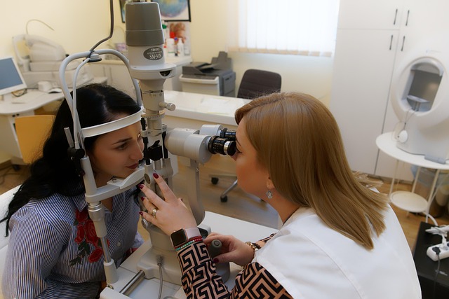
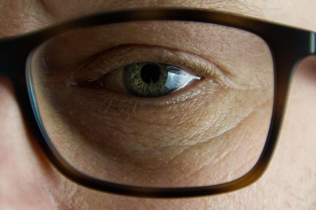
The Turkish team examined the corneal endothelium, which keeps the cornea clear and thin. These cells pump fluid out of the cornea. Endothelial cells do not normally divide in adult eyes. Loss can therefore accumulate over the years. The researchers scanned both corneas before the first dose, and they scanned again two months after the second dose. The vaccine studied was the Pfizer COVID-19 Vaccine, also called BNT162b2. So, what did they discover? Well, the paper’s conclusion is explicit and cautious.
It states, “Changes in corneal endothelium occur in the short term after two doses of the Pfizer-BioNTech (BNT162b2) COVID-19 mRNA vaccine.” It also states, “Hence the endothelium should be closely monitored in those with a low endothelial count or who have had a corneal graft.” Those quotes reflect a measured clinical message. They state that we should pay close attention to those with eyes most at risk. Additionally, they should keep normal eyes under routine care. Lastly, they should continue surveillance to see if changes persist or resolve.
The Potential Consequences of Corneal Endothelial Change
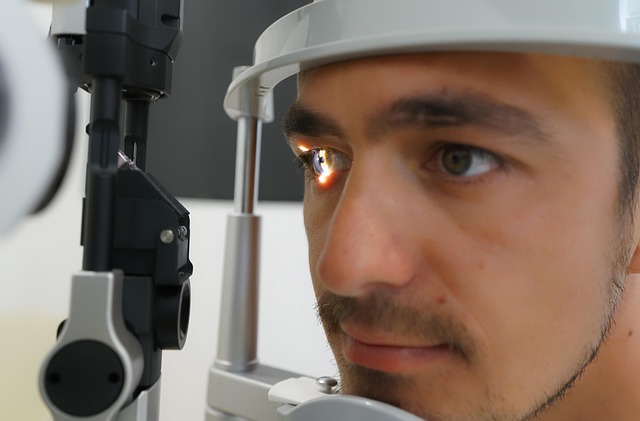
The corneal endothelium works like tiny pumps that keep the cornea clear. When too many cells are lost, fluid builds up and swelling follows. Early stress can show as extra thickness or uneven cell shapes. Doctors call that pleomorphism, and they watch it closely. They use slit lamp exams and specular microscopy to check progress. The American Academy of Ophthalmology explains how this layer controls hydration. Those pumps help you see clearly during normal daily life. Small shifts can be silent at first, so people feel fine. Bigger or lingering shifts can bring blur or discomfort. People with low starting cell counts have less safety margin. Patients with corneal grafts can tip into trouble faster. That is why doctors keep the closest eye on those groups.
In the Turkish cohort, no one had sudden, sight-threatening events. There were no urgent emergencies during the short follow up. The scans showed structural changes that deserve longer tracking. The paper does not argue against the Pfizer COVID-19 Vaccine. It asks for closer monitoring in eyes with lower reserve. The study is modest and comes from a single center. There was no unvaccinated comparison group in this analysis. Follow up was short, so we cannot judge durability. These limits are common in early ophthalmic research. Better studies need matched controls and more time. Teams should also agree on clear thresholds before starting. Those thresholds help separate normal noise from true risk over time.
Results of the Systematic Review
A separate paper reviewed ocular problems reported after COVID vaccination. It covered 20 peer-reviewed studies and 243 patients. Ages ranged from 18 to 84 years. The review states, “The most common ocular complications were ocular inflammatory complications.” It also states, “Despite the high importance of the COVID-19 vaccination, it was found that it is associated with the occurrence of some ocular complications.” The review counted herpetic eye disease across several reports. It counted optic neuritis in a substantial minority. It listed retinal vascular events in a smaller share. The review did not claim proof of causality. It called for better-designed studies to estimate risk. It also highlighted the known ocular effects during COVID infection itself. Many reported problems respond to timely anti-inflammatory care. That remains standard ophthalmic practice.
How Researchers Interpret Rare Ocular Events
Case reports and small series help doctors notice unusual patterns early. They do not tell us how common a problem is. That is why big safety systems run alongside daily care. In Australia, reports go into national checks that scan for signals. A single report does not prove cause, but it can start a review. South Africa uses a similar approach across primary clinics and hospitals. In the United States, linked records power near real-time checks. The Vaccine Safety Datalink looks for spikes and compares groups. VAERS helps spot early patterns across many products. These tools guide labels and clinical advice when signals grow. They also help show when early worries do not hold up.
Doctors also look at what the infection does to the eye. Reviews describe eye surface problems and deeper retinal issues during COVID disease. That history shapes how people weigh vaccination choices today. Some studies disagree about retinal vessel blockages after vaccination. Authors therefore ask for caution with those mixed results. The larger review still finds these outcomes are uncommon. Inflammatory problems make up the biggest share of reports. Overall, the message is careful and steady. Infection is not harmless for the eye. Vaccination can lower severe inflammation during waves. That may reduce some later eye risks. Decisions should consider local spread and personal risk. They should also reflect the person’s eye history and current reserve.
Practical Guidance
Most vaccinated people will not have any eye issues. However, some patients may require special planning with their ophthalmologists. Corneal graft recipients should book a proactive follow-up after vaccination. Patients with very low endothelial counts should also plan ahead. Those patients may already be monitoring their intraocular pressure and corneal thickness. Baseline scans can help doctors compare later measurements with confidence. Furthermore, cataract candidates with borderline counts can discuss timing options. They can space surgeries and follow up with corneal imaging. Anyone with persistent blur, pain, or light sensitivity should seek care. Early anti-inflammatory treatment can prevent long recoveries. National regulators advise that most post-vaccine events are mild. They also advise prompt reporting of anything unusual or severe. That feedback improves surveillance and clinical guidance for everyone.
What Better Studies must still Answer
Big questions usually shrink when studies get larger and run longer. That is the plan here as well. Researchers need comparison groups from similar clinics, so differences are fair. They also need shared protocols that say exactly how to scan and when. Careful records of infections and reinfections will matter a lot. That detail helps separate vaccine signals from infection signals. Teams across hospitals can pool patients who have grafts or low counts. They can also compare how people do when surgeries happen at different times. Linked health records can then catch emergency visits and new diagnoses. They can also show which medicines were used and how patients responded.
All of those steps point to the same goal, which is clarity. We want to know which corneal changes last, and which fade without trouble. Clear answers will guide labels, clinic advice, and follow-up plans for higher-risk eyes. To get there, researchers should agree in advance on meaningful change. Independent graders should read images without seeing earlier results. Every site should use the same equipment and settings, and they should track corneal thickness with the same scans. Results should be adjusted for age, diabetes, and prior eye surgery, so the comparisons stay fair and useful.
What Pfizer has Said in Response
Pfizer has addressed media questions about ocular reports. The company’s statement is cautious and standard for safety. It says, “Patient safety is paramount and we take any reports of adverse events very seriously.” It continues, “Adverse event reports do not imply causality, and in the context of vaccination, such events may be unrelated to administration of the vaccine.” Pfizer also notes the global number of doses administered. It states that the overall benefit-risk profile remains positive. It describes ongoing pharmacovigilance in partnership with regulators. It encourages anyone with concerns to speak with clinicians. That includes pharmacists, nurses, and doctors. These points match regulator guidance about safety signals. They also match the usual language for risk communication.
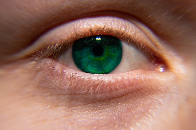
Corneal Biology
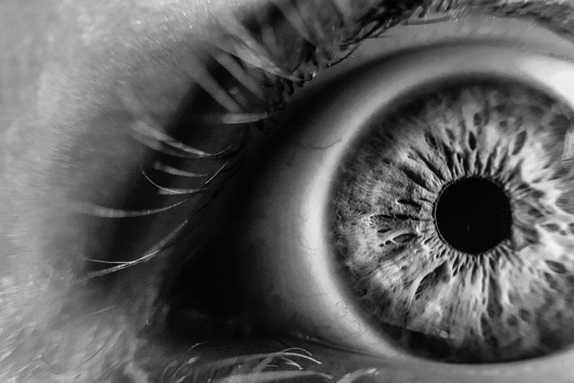
Think of the cornea as a clear window at the front of the eye. On its inner surface sits the endothelium, a single, very thin cell layer. These cells act like tiny pumps that move fluid out of the cornea. When the pumps work well, the window stays clear and vision stays sharp. However, if the pumps slow down, extra fluid lingers in the tissue. The cornea then swells, and the window can look a little foggy. People may notice halos around lights or blurred vision when they wake. Additionally, some feel discomfort, which can show up as ache or light sensitivity. Eye doctors track two simple signals on special scans. “Hexagonality” describes how neatly the cells fit together.
Healthy cells look uniform, and they pack like tiles on a floor. “Cell density” is the count of those cells in a tiny area. Falling density or messy shapes suggest the layer is under stress. Short dips can settle with time and routine follow up, whereas longer dips can leave the cornea with less safety margin. That lower reserve makes future stressors more risky for vision. This is why the Turkish team urged closer monitoring. They focused on people with a corneal graft or very low counts. The authors did not tell anyone to stop getting vaccinated. They asked for more research and sensible, careful follow up. If someone has a vulnerable cornea, a planned check can help. Baseline scans and a quick review after shots often answer questions. That simple plan keeps problems small and helps protect everyday vision.
Putting the Two Papers in one Frame

The Turkish cohort studied structure with paired measurements. It flagged short-term endothelial shifts after vaccination. It also flagged a narrow group for closer follow-up. The systematic review surveyed published cases. It tallied mostly inflammatory problems across reports. It included herpetic disease and optic neuritis, among others. It also counted retinal vascular events in a smaller share. The authors called for better designs that estimate risk. They stressed careful interpretation of case-based evidence. The two papers, therefore, answer different questions.
One addresses corneal structure soon after vaccination. One summarizes rare events reported in many settings. Read together, they support sensible vigilance and continued research. They do not justify fear or complacency. Importantly, the cohort tracked paired eyes within individuals, improving comparability. Its caution focuses on patients with limited endothelial reserve. The review aggregates signals across settings, not incidence estimates. As the reviewers note, “The most common ocular complications were ocular inflammatory complications.” Together, these approaches map signals and guide smarter, targeted monitoring.
Read More: Father Shares Experience of Illness Following Pfizer COVID Vaccine, Discusses Recovery
How Clinicians can Apply this Today
Eye teams can prepare simple playbooks for higher-risk patients. They can time specular microscopy before and after doses. They can schedule earlier reviews for graft recipients. They can encourage reporting of persistent symptoms after vaccination. They can also place ocular risk in the context of infection risk. Many clinics already use these workflows for surgery planning. The same approach fits vaccine follow-up for vulnerable corneas. Primary care teams can coordinate with ophthalmology when needed. Pharmacists can encourage prompt care for enduring symptoms.
National systems explain that most events are mild and short. They also explain how to report anything significant. This shared approach balances prudence and access. It supports safety without discouraging needed protection. Clinics can add a short checklist to post-vaccine discharge notes. It can list red flags, contact numbers, and follow up windows. Electronic records can flag patients with grafts or low counts. That flag prompts schedulers to book earlier reviews automatically. Community optometrists can share specular images securely with surgeons. That simple pipeline speeds triage.
The Bottom Line
Two conclusions stand on the evidence today. First, the Turkish cohort reports short-term corneal endothelial changes after two doses of the Pfizer COVID-19 Vaccine. The authors write, “Changes in corneal endothelium occur in the short term” and “Hence the endothelium should be closely monitored” in lower-reserve eyes. Second, the systematic review catalogs rare ocular problems after COVID vaccination. It states, “The most common ocular complications were ocular inflammatory complications.” It also states, “it is associated with the occurrence of some ocular complications.”
Basically, these papers do not indicate any widespread vision loss from vaccination. They support targeted vigilance and continued research. Regulators still judge the overall benefits of vaccination to be favorable. However, people with grafts or very low counts should plan a closer follow-up. Anyone with persistent blur, pain, or light sensitivity should seek care. Shared decisions can then reflect personal risks and local transmission. For context, regulators also stress report interpretation. The TGA says, “The fact that an adverse medical event… does not mean that the vaccine has caused it.” That message supports meticulous study while avoiding premature conclusions.
Disclaimer: This article was created with AI assistance and edited by a human for accuracy and clarity.
Read More: What is Vizz? The New FDA-Approved Eye Drop to Fix Near Vision