While it was long-believed that our chromosomes determined an embryo’s gender from the minute sperm impregnated the egg. However, new research may contest these previous findings. Researchers from Osaka University conducted a study that reveals how low iron during pregnancy could potentially influence fetal gender determination. The team discovered that low iron during a pregnancy can influence genetic gender, changing male (XY) embryos into females (XX).
However, these findings were discovered in animal trials. Human trials still need to be conducted but the results are promising. Only around week 7 does the developing embryo switch to a deterministic biological program to develop testes or ovaries. This has raised interesting questions like the influence of nutrient deficiencies on genetic gender programming.
Iron Deficiency and Gender Development
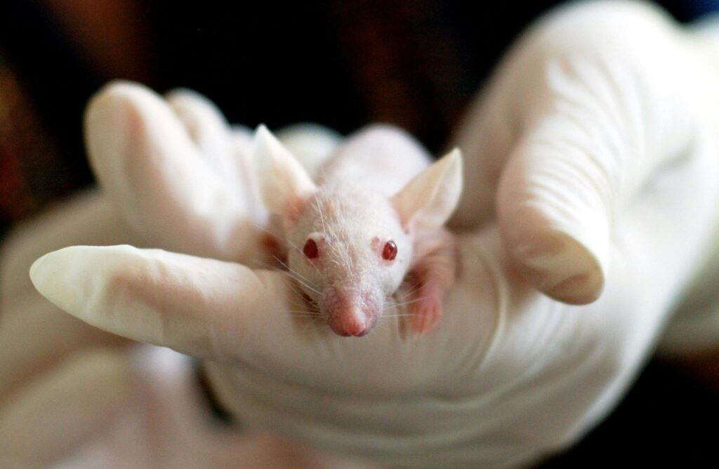
Scientists at Osaka University discovered that iron deficiency during pregnancy can cause male mouse embryos to develop into females. This demonstrates that 6 out of 39 genetically male mice (XY) developed ovaries when their mothers experienced iron deficiency. The mechanism involves a crucial enzyme called KDM3A, which requires iron to function properly. This enzyme activates the SRY gene, which is responsible for testes formation. When iron levels drop, KDM3A cannot remove the chemical blocks that keep the SRY gene silenced, leading to female development instead of male development.
Read More: It’s Officially Time to Retire the ‘Gender Reveal Party’
The Science Behind Gender Development Changes

The study reinforced their findings that iron deficiency disrupts the epigenetic regulation of SRY gene activation. The SRY gene on the Y chromosome typically triggers testis formation, but it requires iron-dependent enzymes to become active. KDM3A, a histone demethylase enzyme, needs ferrous iron to activate the SRY gene; without it, the SRY gene remains dormant. If the SRY gene is not activated, ovaries will form by default.
Testing the Impact Of Iron
Researchers used multiple experiments to conduct their idea. In one experiment, researchers removed iron from lab-grown gonads of XY embryos. This caused the SRY gene to turn off and cells to show ovarian markers. Gonads are the glands responsible for the production of gender-related hormones.
Afterwards, they disabled the genes favoring iron accumulation in these embryonic gonads. Among 39 genetically male embryos, 6 grew ovaries while 1 developed mixed types of organs. This meant nearly 1 in 5 showed complete or partial gender changes.
In their second experiment a pharmaceutical was introduced to pregnant mice that diminishes iron. Among the 41 XY embryos, 6 developed 2 ovaries while 3 developed mixed gonads. Scientists also tested if diet alone could trigger these changes. They fed pregnant mice a low-iron diet and only mice with a gene mutation in the Kdm3a gene developed ovaries. 2 out of 43 male embryos developed female organs, proving iron’s role in gender development.
Timing and Critical Windows
The research identified a remarkably narrow window of vulnerability. Gender determination in mammals occurs during approximately 48 hours when embryonic gonads decide their fate. Missing iron delivery during this brief period can permanently switch genetic males toward female development.
Broader Health Implications
This research does give valuable insight into gender determination but may also prove beneficial for broader, serious health complications. Low iron during pregnancy significantly increases risks for both mothers and babies, including preterm delivery, low birth weight, and postpartum complications. Maternal iron deficiency anemia more than doubles the odds of preterm delivery and triples the risk of low birth weight.
Iron Requirements and Supplementation
Pregnant women need approximately 27 mg of elemental iron daily, to prevent increased risk of anemia, infection or in worst cases, misscarriage. The World Health Organization recommends daily supplementation with 30-60 mg of elemental iron during pregnancy, with higher doses preferred in settings where anemia is a severe public health problem.

Most prenatal vitamins contain about 30 mg of iron, but women with existing deficiency may need higher doses. The iron required during pregnancy drastically increases from 1.0 mg per day in the first trimester to 7.5 mg per day in the third trimester. It is common, especially during the third trimester, for many women to suffer from an iron deficiency.
Postpartum Consequences
Iron deficiency can continue to impact mothers after delivery. Postpartum anemia affects 22% to 50% of women in developed countries, primarily due to untreated iron deficiency and peripartum blood loss. This condition increases risks of fatigue, depression, impaired cognition, and could negatively impact a mother bonding with their new born.
Research shows that iron deficiency anemia independently increases the risk of atonic postpartum hemorrhage by 58.8%. Women with iron deficiency anemia have significantly higher rates of total postpartum hemorrhage (4.5% versus 3.2%) and atonic hemorrhage (3.1% versus 2.0%).
Future Research Directions
The Osaka University findings open entirely new research avenues. While the gender determination effects have only been demonstrated in mice, the implications warrant investigation in human populations. Professor Peter Koopman from the University of Queensland noted that “iron deficiency affects an estimated 40% of pregnant women, and iron has not been previously suspected of influencing gender determination in the embryo.” While these findings are fascinating, emphasis on global iron deficiencies in women during pregnancy should be addressed.
Read More: Leaked Documents Stoke Fears About Gender Treatment in Children
